Physicians
have been largely silent on two
nuclear industry challenges before the federal government. Do
we pour millions of dollars into research and development of Small
Modular Nuclear Reactors? Do
we bury our current nuclear waste in the vicinity of Wakerton,
Teeswater and Kincardine and
the city of Ottawa in
Ontario?i
In fact, physicians have been silent about nuclear power in general. We have also been silenced as the industry has worked its mysterious media fantasy of “too cheap to monitor” into a myth of “necessity” for climate change energy. We have been silent, not because we have nothing to say but because we’ve been led to believe that these decisions are “political”, not health-related.
During the Canadian Covid epidemic, physicians were utterly unable to keep the primacy of health care out of the political arena. We saw our ICUs worked beyond capacity, and our colleagues quit in frustration. With respect to the threat of ionizing radiation we have been remarkably silent.
To reiterate, the two very different questions currently before Canadian legislators, both involve ionizing radiation, not merely that of nature (sun, rocks, air) but entirely man-made atoms, some of which will still be emitting ionizing radiation in a time-frame that is outside human comprehension (plutonium-239 will be around for 24,100 x 10 yearsii).
1. Nuclear Waste Management:
Having decided that the only way to “manage” nuclear waste was to put it into a Deep Geological Repository (DGR), the Nuclear Waste Management Organization (NWMO)iii has spent the last two of decades searching for such a site. The process has divided communities even while much of negotiation with perceived leaders (mayors, chiefs and council members) has occurred behind closed doors. Health professionals have been silent.
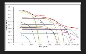
These wastes will be toxic for a long, long time. They are not like ordinary waste simply degrading over time. Each nuclear element has its own decay chain. What is buried in 2030 will never be what it was again. Short-lived waste and decay products will disappear but longer-lived ones will continue to emit radioactivity. A cask of nuclear waste is like a boiling cauldron with atoms of ever-changing elements.
From this graph, you can see that the total radiation has decreased over 10 million years.
While it is easy to understand that burying the waste in undisturbed bedrock changes the bedrock to a “disturbed” status, many people don’t know that the radioactivity of the waste can change the very containers in which they are buried. Refurbishing of nuclear reactors involves replacement of miles of metal pipes that have become corroded, why would we expect that containers in a DGR would fare better?iv
DGR? This is not the only disposal in question – NWMO is fantasizing a Near Surface Disposal Facilityv (NSDF) at Chalk River only 200 km North of Ottawa. This proposed disposal facility would be an eight story mound that will be within a few hundred metres of the river from which Ottawa and Montreal draw their cities’ drinking water.
vi From
Ionizing radiation directly affects the human genome. No one disputes this but many believe or pretend to believe (denial) that there is a low level at which it is actually beneficial. Hormesis is a fiction.
Physicians often find themselves at odds with an industry when, using the precautionary principle, we hesitate to approve a new drug or technical procedure. For example, thalidomide’s disastrous side effects became evident only after marketing. Our history with radiation in medical use has been a continual story of overuse followed by rationalizing behaviour.
A radiological example, X-rays and CT scans. The overuse of x-rays for removal of skin lesions led to discipline by the American Medical Association at the end of the 1920’s and use for hair removal forbidden. Using x-rays to treat Tinea capitus in Israel resulted in a significant increase in cancers of the head and neck decades later.
In 1996, I joined the multitude of physicians using CT scans “to rule out brain injuries” in children that had bonked their heads – a negative result tended to reassure both the parents and myself, only to discover that these investigations resulted in an increase in cancers years later.vii Choosing Wisely, a program to guide physician decisions about lab and x-ray investigations, now recommends CT scans under a limited number of circumstances – none of which are “I want reassurance” or the “parents expect it.
The industry has settled upon a Deep Geological Repository (DGR). What could be simpler? Bury it deeply in the bedrock where it would be safe and immobile for time immemorial. Are we so brilliant and so prescient that we can know what will be safe for the next 100,000 years and more?
Currently the waste is stored in concrete containers above ground. They can be watched – providing jobs for generations of Canadians – and repackaged if they leak. “Rolling Stewardship” would provide laboratories in Canada and elsewhere with experimental material. Finally, perhaps our brighter descendants will discover a way to use ionizing radiation safely. Instead of choosing a DGR recklessly with our limited understanding of the waste, we have the opportunity to spawn years of research.
2. Building Small Modular Nuclear Reactors:
Full page ads about Small Modular Nuclear Reactors tout them as “safe”. How is anything that produces ionizing radiation safe? We use it extensively in diagnostics and treatment but we also know that it is implicated in causing cancer.
In the 1950’s we routinely x-rayed pregnant women for “pelvimetry”, measuring their bony pelvic outlet to ascertain whether they could give birth naturally or needed a Caesarian section. In both the USA and the UK, the cancer societies noted a rise in leukemia in children. Suddenly there was a boom in building pediatric hospitals devoted to treating cancer.
During the 1940’s and 1950’s, there was a leukemia boom in North Armerica and the UK. There was a sudden increase in incidence from ~ 1:20 to 1:16 children. At that time it was almost always fatal.
Dr. Alice Stewart, a general practitioner in the UK and one of the first epidemiologists, and the Tri-State Health Study in the USA collected data and connected the dots. X-rays during fetal development doubled the incidence of cancer in the offspring. Doctors had been ordering x-rays to measure womens’ pelvises, a series called “”pelvimetry”. The technique disappeared. Lead aprons came out for dental x-rays.
In 2007, after decades of study and six reports, the Biological Effects of Ionizing Radiation (BEIR VII)viii, in publishing its seventh report concluded that any level of exposure to radiation was unsafe (although “at lose doses, the number of radiation induced cancers is small”).
Studies of significant increases in leukemia and other cancers within 5 to 25 km of operating nuclear power plants seem to yield conclusive results but research by both the Committee on Medical Aspects of Radiation in the Environment (COMARE, UK) and the Kikki study in Germany have had disputed by the nuclear industry.
iP. Kaatsch, C.Spix, S. Schmiedel, R. Schulze-Rath, A.Mergenthaler, and M. Blettner, Epidemiologische Studie zu Kinderkrebs in der Umgebung vonKerkraftwerken (KiKK Studie), Sltzgitter: Bundesamp fuer Strablenschutz, 2007, urn:nbn:de:0221-20100317939.
In 2012, I mentioned the German study in discussion after delivering a brief to the Canadian Nuclear Safety Commission (CNSC). The health science person on the board dismissively called the effect “due to a virus.” If there is a special virus that affects only children close to nuclear power plants, we should endeavour to identify it. No such research appears to have been launched.
Before 1990, when it was forced to open its records, the United States Department of Energy not only controlled access to all information on the health effects of radiation on nuclear workers and the public but it also controlled all the funding of radiation researchx. It had successfully stone-walled independent research for decades.
We don’t know which ray or particle will cause any particular DNA molecule to turn the cell into cancer but we know that they do. Physicians have conducted studies on the medical uses of ionizing radiation. Here are a few examples:
a) Xray treatment of Tinea capitus:
The studies on patients who, as children, received x-ray treatment for Tinea capitis (fungal infection of the scalp) in the 1940’s and 1950’s have shown “excess incidences of tumours of the head and neck including the skin, brain, thyroid, and parotid glands”xi. Needless to say, this technique of treating fungal infections is left in the dustbin of history.
b) Breast cancer after fluoroscopic examinations of the breast during treatment for pulmonary tuberculosis:
“The role of ionizing radiation as a cause of carcinoma has long been recognized, particularly in relationship to carcinoma of the skin, lung, thyroid and bone.”xii Dr. Ian Mackenzie in Halifax found fifty cases of breast cancer in patients who had received this form of x-ray treatment prior to 1961. From length of time from the beginning of the x-ray treatments and the breast cancer averaged 17 years. There was also a high correlation between the side of treatment and the cancer-affected breast.
This method of treating pulmonary TB had disappeared by 1955 when antibiotics became available but This research carried out largely on indigenous women and largely had disappeared by 1955. Decades later when they developed breast cancer, I’m sure they were very grateful that they had contributed to show that breast tissue was sensitive to radiation. Should we take a closer look at the use of x-rays in mammograms?
c) PET, MUGA, MIBI and SPECT scans:
All of these scans use a radioisotope, an element that gives off gamma rays that can create images of various parts of the body. For example, I131 concentrates within 15 minutes in the thyroid. This can provide a very good picture of the thyroid. Some studies require two scans, one before the radioisotope is injected and then a later one showing where the element accumulates in the body.
In 2011 researchers in Montreal examined the charts of more than 80,000 patients who had received post-heart attack PET scans and concluded that there was a 3% increase in the incidence of cancer per 10 mSv of scan exposurexiii. Scans require between two and 8 exposures to satisfy the demands of the test. (For comparison, 10 mSv is equivalent to the exposure from 100 chest x-rays).
d) Increased secondary cancers in post-radiation patients:
“Radiation-induced second malignancies (RISM) is one of the important late side effects of radiation therapy”.xiv The exact risk is dependent upon so many factorsxv that it probably contributes only 5% increase of the 17 – 19% total secondary cancers.
What is quite amazing is the real paucity of good prospective research on the health effects of radiation. These ones briefly listed here concentrate on carcinogenic effects, there are other possible affects. Radiation-associated cardiac diseasesxvi is known. Hypertension with its related cardiovascular diseases and strokes has also been identified as associated with chronic exposure to low-dose radiationxvii.
We know that background radiation has health effectsxviii but it seems that limited research has been conducted on diseases other than cancer.
Many of the residents of Port Hope, home to Canada’s uranium refinery since 1933, feel that they have been “researched to death”xix but a casual review of papers shows that many have time-lines that are too short, populations sizes that are too small, and mixed outcomes which do a disservice to all. Quantity of research tells us nothing if it's poorly done.
Finally, the myth believed by many physicians and the public is that we need nuclear power for radioisotopes, for treatment or diagnosis. We do not. We already make radioisotopes more safely in cyclotrons or accelerators and could expand this to all medical radioisotopes.
In conclusion, ionizing radiation is not safe; nuclear power cannot be made safe. Physicians have been altogether too silent about this industry – or, in fact, silenced at its very inception. With the pressure on the government to support an industry that is too slow, too costly and too dangerous to respond to the threatening climate change, who speaks for our great grandchildren?
iBurying nuclear waste requires resources and uses energy as well – currently high grade steel containers covered in copper have been found to be the least likely to corrode. These do not seem to be costed out in either monetary or environmental terms.
iiThe half-life of plutonium-239 is 24,900 years; it takes ten half-lives to disintegrate to almost unmeasurable amounts.
iiiThe Nuclear Waste Management Organization (NWMO, pronounced “Noo-mo”) was formed in 2002 by Canada’s nuclear electrical energy providers as directed by the Nuclear Fuel Waste Act (NFWA). https://nwmo.ca
ivNWMO engineers say that the containers won’t corrode because they are made of the “finest steel” and covered with relatively impervious-to-radiation copper. Does this mean that the tubing in nuciear reactors is not the “finest steel”?
vhttps://www.theglobeandmail.com/canada/article-canada-nuclear-waste-management/
viFrom Geosphere: A blog hosted by the European Geoscience Union. Note that the graph starts at a “zero” of 1000 Bq. https://blogs.egu.eu/network/geosphere/files/2014/12/Untitled.png
viihttps://www.choosingwisely.org/clinician-lists/american-academy-pediatrics-ct-scans-to-evaluate-minor-head-injuries/
viiihttps://nap.nationalacademies.org/resource/11340/beir_vii_final.pdf
ixP. Kaatsch, C.Spix, S. Schmiedel, R. Schulze-Rath, A.Mergenthaler, and M. Blettner, Epidemiologische Studie zu Kinderkrebs in der Umgebung vonKerkraftwerken (KiKK Studie), Sltzgitter: Bundesamp fuer Strablenschutz, 2007, urn:nbn:de:0221-20100317939.
xhttps://www.sfgate.com/bayarea/article/Alice-Stewart-her-research-led-to-end-of-2800048.php
xiFollow-up study of patients treated by X-ray epilation for Tinea capitis: https://pubmed.ncbi.nlm.nih.gov/1244805/
This paper is merely one of many on this subject.
xii J. A. Myrden, J. E. Hiltz, “Breast Cancer Following Multiple Fluoroscopies During Artificial Pneumothorax Treatment of Pulmonary Tuberculosis”, Canad. Med. Ass.J., June 14, 1969, vol 100
xiiiMark J. Eisenberg, Johathan Afilalo, Patrick R. Lawler, Michal Abramhamowicz, Hugues Righard, and Louise Pilot, “Cancr Risk Related to Low-Dose Ionizing Radiation from Cardiac Imaging in Patients after Acute Myocardial Infarction”, Canad. Med. Ass. J, 183(March 8, 2011); 430-436.
xivChinna Babu Dracham, Abhash Shankar, and Renu Madan,”Radiation induced secondary malignancies: a review article” Radiat Oncol J. 2018 Jun; 36(2): 85–94. https://www.ncbi.nlm.nih.gov/pmc/articles/PMC6074073/
xv For example: age at radiation, dose and volume of irradiated area, type of irradiated organ and tissue,
radiation technique, individual and family history of cancer, chemotherapy, cigarette-smoking, diet and
external environment.
xviMilind Y. Desai, “Radiation Associated Cardiac Disease”, American College of Cardiology, June 21, 2017. https://www.acc.org/latest-in-cardiology/articles/2017/06/13/07/13/radiation-associated-cardiac-disease
xviihttps://www.heart.org/en/news/2019/05/03/regular-low-level-radiation-exposure-raises-high-blood-pressure-risk
xviiiBen Spycher, Judith Lupatsch, Marcel Zwahlen, Martin Roosli, Felix Niggli, Michael Grotzer, Johannes Rischewski, Matthias Egger, Claudia Kuehni, for the Swiss Pediatric Oncology Group and the Swiss national Cohort Study Group, “Background Ionizing Radiation and the Risk of Chilhood Cancer: A Census-Based Nationwide Cohort Study”, Environmental Health Perspectives, June 1, 2015. https://ehp.niehs.nih.gov/doi/full/10.1289/ehp.1408548
